Objective:
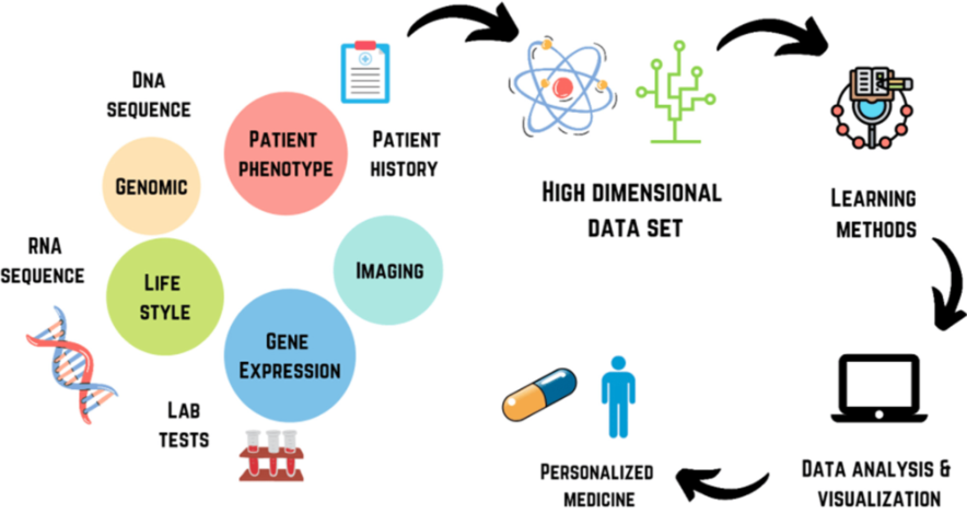
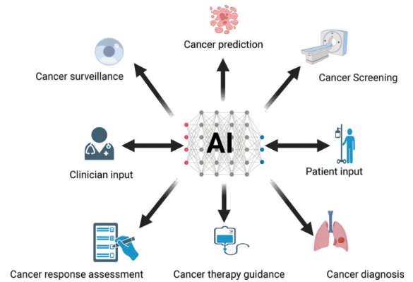
Develop an integrated AI model that utilizes clinical, pathological, genomic data, and image analysis for accurate prediction of oncology treatment success rates and cancer staging. This AI cancer treatment prediction model enables faster and more accurate diagnosis for cancer stages, providing beneficial insights and predictions towards treatment procedures. Our aim is to develop a flexible and robust model that is updated with global health trends and used as an analysis tool by professionals.

Development Cycle:
It is of utmost importance that accurate feature engineering is undertaken to provide more accurate co-relations between different factors. Which in turn would enable more accurate predictions by the model.
Following are the steps undertaken for development of treatment success rate predictive model:
1) Data Collection: Aggregate diverse datasets including clinical records, pathology reports, and genomic profiles of cancer patients.
2) Feature Engineering: Extract relevant features from clinical, pathological, and genomic data to create a comprehensive dataset for model training.
3) Data Pre-processing: Address missing values, standardize formats, and normalize data for consistency.
4) Treatment Success Prediction Model: Develop a machine learning model, possibly a combination of regression and classification algorithms, to predict the success rate of different oncology treatments.
5) Integration of Data: Fuse clinical parameters, pathological findings, and genomic information to enhance the model’s predictive capabilities.
6) Outcome: Provide clinicians with a tool for personalized treatment recommendations based on individual patient profiles.
Steps undertaken for development of cancer stage predictive model:
STEP 1
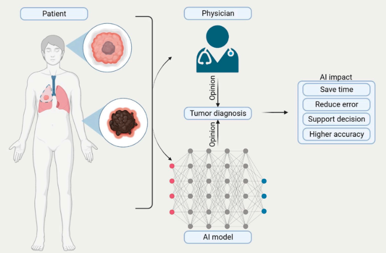
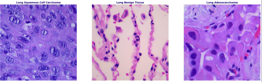
Image Analysis for Cancer Staging: Implement deep learning algorithms to analyse microscopic images of cancer cells and pathological features.
STEP 2
Dataset Labelling: Creating a labelled dataset from microscopic images with different classes based off the morphological factors of the mutated cell.
STEP 3
Stage Prediction Model: Train a model that predicts cancer stage based on the characteristics identified in the microscopic images.
STEP 4
Integration with Clinical Data: Combine image-derived information with clinical, pathological, and genomic data to refine cancer staging predictions
Key Features:
Interdisciplinary Integration: Leverage insights from clinical, pathological, genomic, and image data to create a holistic understanding of each patient’s cancer profile.
Real-time Analysis: Develop algorithms capable of real-time analysis, allowing for swift decision-making and timely interventions.
User-Friendly Interface: Create an intuitive user interface for healthcare professionals, presenting comprehensive reports that incorporate all relevant data for informed decision-making.
Expected Impact:
- Improved Treatment Outcomes: Enhance treatment success rates by tailoring therapeutic approaches based on a patient’s unique clinical, pathological, genomic, and imaging profile.
- Efficient Cancer Staging: Facilitate accurate and timely cancer staging through automated analysis of microscopic images, aiding in treatment planning.
- Evaluation: Assess the model’s performance through rigorous validation using diverse datasets and real-world clinical scenarios.
- Advancement in Personalized Medicine: Contribute to the paradigm shift towards personalized medicine in oncology, optimizing patient care and resource allocation.
- Future Directions: Continuously update the model with new data to improve its predictive accuracy. Explore opportunities for integration with electronic health records (EHR) systems for seamless adoption in clinical settings.